What is Exposure Keratopathy ? |
Exposure keratopathy refers to that surface disorder of the eye in which absence of an adequate tear film results in breakdown of the corneal epithelium. This may produce disruption of corneal epithelium, ulcer formation, superadded infection, thinning and even perforation of eyes in severe cases.
Exposure keratopathy is broadly divided into two main categories viz.
- Exposure due to primary or secondary corneal anaesthesia or hypoaesthesia: Corneal sensation plays an important role in the maintenance of healthy corneal epithelium. It is presumed that nerve growth factors released by corneal nerves mediate epithelial cell proliferation. Thus, corneal anaesthesia results in a surface which is more vulnerable to injury, delayed healing of corneal epithelium, and progressive keratopathy. Reduced corneal sensation may be congenital or acquired, and may be complete or partial.
- Exposure produced by mechanical eyelid abnormalities such as lagophthalmos: Lagophthalmos is the inability to close eyelids completely. Lagophthalmos may be divided into three main groups e.g. Proptosis causing excessive ocular surface exposure, palpebral (eyelid) insufficiency secondary to physiologic, congenital or acquired conditions, and idiopathic lagophthalmos. Lagophthalmos with or without exposure keratopathy may be present at night in some healthy individuals (nocturnal lagophthalmos).
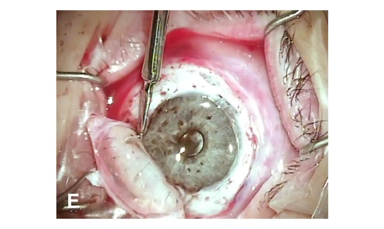
Exposure of cornea from any cause may lead to thickening, xerosis (dryness), and scarring of epidermis
Exposure Keratopathy Symptoms |
- Irritation of eyes.
- Foreign body sensation.
- Burning.
- Blurring of vision.
- Watering.
- Redness.
- Photophobia (increased sensitivity to light).
- Corneal vascularisation.
- Corneal ulceration.
Exposure Keratopathy Causes |
I. Neurotrophic
- Fifth intracranial nerve palsy.
- Cerebro-vascular accident.
- Aneurysm.
- Tumours.
- Herpes simplex.
- Herpes zoster.
- Multiple sclerosis.
II. Neuroparalytic
- Intracranial seventh nerve palsy.
III. Malposition of lids
- Lagophthalmos.
- Proptosis.
Exposure Keratopathy Diagnosis |
Clinical history: To determine the aetiology, history should elucidate any recent trauma, surgery, or any infection. History of Bell’s palsy should be taken. A family history of corneal anaesthesia or lagophthalmos should be elicited.
Clinical examination: The position of eyelids in a relaxed open and closed position as well as inter-palpebral distance should be assessed. Appositional closure of the eyelids with each blink should be recorded. Slit-lamp examination may be done to exclude any obscure lagophthalmos. Obscure lagophthalmos may be due to when the upper and lower eyelashes meet, preventing complete closure of the lids, or obscures view of the true eyelid position. The upper eyelid may also overhang the lower lid, giving the appearance of total closure, however, eyelid margins may not oppose. Bell’s phenomenon should be recorded.
Conjunctiva is examined for any areas of scarring or cicatrisation.
Corneal sensations are evaluated by using wisp of cotton or an aesthesiometer.
Staining with Fluorescein sodium dye may reveal any punctuate epithelial erosions, frank epithelial defects, or ulceration.
A complete neurological examination is also conducted including assessment of cranial nerves.