What is Atopic Keratoconjunctivitis ?
Atopic keratoconjunctivitis (AKC) is a relatively uncommon, but potentially blinding ocular (eye) disease, which typically affects young people with atopic dermatitis. AKC is a severe chronic inflammatory disease of the conjunctiva which may have dramatic consequences for visual function.
The incidence of atopic dermatitis is greatest in paediatric population. AKC may occur at any time after the onset of the associated dermatitis or other atopic condition, and is not necessarily correlated with exacerbations of these conditions. The initial presentation of ocular symptoms in AKC most commonly occurs in the second to third decade of life, with some patients presenting earlier or later (range varies from late teens to 50 years with peak age of incidence in people aged 30-50 years). Visually significant complications most frequently occur in the fourth to fifth decades, with more men affected than women. The condition then remains chronic for years, usually requiring lifelong treatment. An earlier onset of AKC carries the greatest risk for tear film abnormalities and greater ocular surface damage. Classically involved skin areas of atopic dermatitis are lateral neck folds, antecubital (in-front of the elbow) and popliteal (back of knee joint) fossae, but may involve other parts of body including eyelid.
In the year 1952, Hogan described bilateral conjunctivitis occurring in patients with atopic dermatitis (coined the term atopic keratoconjunctivitis), which flared-up with worsening dermatitis. Patients notably showed chronic conjunctival hyperaemia and thickening, corneal epitheliopathy, and later corneal scarring and neovascularisation. Atopic keratoconjunctivitis may evolve independent of dermatitis in some patients. Hogan’s criteria to aid in diagnosis is presence or history of eczematous dermatitis, a family history of atopic disease, associated allergies (asthma, hay fever), eosinophilia and a characteristic keratoconjunctivitis associated with exacerbations of dermatitis. His patients were notable for chronic conjunctival hyperaemia and thickening, corneal epitheliopathy, and later corneal scarring and neovascularisation.
Atopic keratoconjunctivitis represents a disease under chronic allergic conjunctivitis (CAC), together with seasonal allergic conjunctivitis (SAC), perennial allergic conjunctivitis (PAC), vernal keratoconjunctivitis (VKC) and, to a certain extent, giant papillary conjunctivitis (GPC). The potential for corneal involvement and opacification increases significantly with the CACs because of the severity and sustained nature of their inflammation. In seasonal and perennial allergic conjunctivitis, conjunctival inflammation is quite mild and corneal involvement is rare. However, clinical and patho-physiological features of AKC are quite different from other allergic conjunctivitis. AKC like vernal keratoconjunctivitis, is a severe inflammatory disease, which may involve cornea and can cause permanent visual impairment. The highest incidence of visual loss is therefore, found in AKC, the most chronic of these disorders.
Chronic allergic conjunctivitis characteristically involves an Immunoglobulin E (IgE) mediated type I hypersensitivity response, manifested by papillary conjunctivitis, with itching as a universal early symptom. Hypersensitivity reactions associated with type I and a prominent T-cell mediated type IV response, along with variety of other inflammatory cell types and cytokines, contribute to the inflammatory changes of conjunctiva and cornea seen in AKC.
AKC and VKC are more frequently associated with eczema and asthma respectively, while SAC and PAC are more frequently associated with rhinitis.
AKC tends to be chronic and unremitting, with a relatively low expectation of eventual resolution, and is associated with significant ocular morbidity. VKC is more frequently seasonal, and is generally worse in the spring season. AKC tends to be perennial and is often worse in winter. Patients are sensitive to a wide range of airborne environmental allergens.
Atopic Keratoconjunctivitis Symptoms
Unlike SAC, symptoms of AKC are usually present year-round, though a significant number of patients may have seasonal exacerbations.
Ocular symptoms of AKC:
- Moderate to severe Itching.
- Tearing of eyes.
- Irritation.
- Burning sensation.
- Dermatitis of eyelids and peri-orbital skin.
- Peri-orbital hyper-pigmentation.
- Chronic eyelid oedema and inflammation.
- Entropion (inward turning of eyelid).
- Ptosis (drooping of upper eyelid).
- Lagophthalmos (incomplete or defective closure of eyelids).
- Madarosis (loss of eyelashes).
- Redness of eyes.
- Stringy mucoid discharge.
- Difficulty in opening the eyes on awakening.
- Discomfort in eyes.
- Photophobia (sensitivity to light).
- Pain.
- Blurring of vision.
Atopic Keratoconjunctivitis Causes
Atopic keratoconjunctivitis is a chronic allergic conjunctivitis and may be due to:
Hypersensitivity response:
All ocular allergic disorders are characterised by a hypersensitivity response, defined as an excessive reaction of the normal immune system, usually by exposure to an inciting antigen.
Type I hypersensitivity, or IgE mediated immediate hypersensitivity, predominates in PAC and SAC, but is also involved in other CACs, including AKC. Serum IgE levels are chronically elevated in AKC patients but their levels are not necessarily correlated to severity of disease. IgE levels in tear samples are also increased in AKC, and correlate with serum IgE levels.
AKC additionally involves a chronic inflammation of the ocular surface; a type IV delayed hypersensitivity response, where an immediate antigen is not easily identified.
Genetic predisposition:
A genetic predisposition combined with antigen sensitisation is suspected in AKC and its associated disease, atopic dermatitis. AKC may, however, represent either a common manifestation end point for a number of abnormal gene processes, or a single gene defect with variable phenotypic expression, modified by other gene polymorphisms and the environment.
Epithelial barrier defect:
More recent studies suggest that an epithelial barrier defect may be responsible rather than a defect in immune-regulatory function.
Atopic (genetic tendency to develop allergic disease) systemic diseases with which AKC may be associated are:
- Dermatitis is most common.
- Eczema.
- Asthma.
- Hay Fever.
- Migraine.
- Urticaria (hives).
- Rhinitis.
- Food allergies (less common).
- Non-hereditary angioedema (less common).
AKC may be associated with other ocular diseases like:
- Keratoconus.
- Anterior sub-capsular cataract.
- Posterior sub-capsular cataract.
- Chronic staphylococcal blepharitis.
- Retinal detachment.
Atopic Keratoconjunctivitis Diagnosis
Diagnosis of AKC is based upon typical clinical features.
Despite facts suggesting immune responses in the pathogenesis of VKC, no clinical or laboratory test has evolved to support the diagnosis in atypical cases or predict the course of disease.
History of atopy in patient or the family, elevated serum level of total and specific IgE, higher number of eosinophils and mast cells, increased level of mediators and favourable response to anti-allergic therapy is observed in AKC.
Ocular clinical features:
Peri-orbital features:
- Dermatitis: Scaling, flaky dermatitis consistent with eczema on peri-orbital skin.
- Hyperpigmentation: Peri-orbital hyper-pigmentation, which may lighten in response to control of inflammation.
- de Hertoghe sign: de Hertoghe sign or absence of lateral eyebrow, is occasionally seen and may be related to chronic eye rubbing.
Eyelids:
- Dermatitis: Scaling, flaky dermatitis with reddened base, consistent with eczema on eyelids.
- Vertical corrugations: Vertical corrugations near medial canthus of upper and lower eyelid may result.
- Fissured eyelid skin: Eyelid skin is often fissured.
- Eyelid margin thickening: Eyelid margins may be thickened, oedematous and hyperaemic.
- Dennie-Morgan lines: Eyelid oedema may lead to Dennie-Morgan lines, single or double creases in the lower eyelid, secondary to chronic eye rubbing.
- Lateral canthal ulceration: Lateral canthal ulceration related to chronic tearing may be present.
- Lid mal-position: Lid mal-position may result from chronic eyelid oedema and inflammation, resulting into:
1. Ectropion (outward turning of eyelid).
2. Ptosis.
3. Lagophthalmos may result from chronic eyelid oedema and inflammation.
- Madarosis: Madarosis may also result from chronic eyelid oedema and inflammation. Lid margins may show loss of cilia and punctal ectropion.
- Permanent lid swelling: Chronic oedema may lead to permanent lid swelling, a hallmark of long-standing atopic eye disease.
- Eyelid margin keratinisation: Keratinisation of eyelid margins is sometimes observed, along with associated meibomitis (inflammation of meibomian glands) and obliteration of meibomian glands.
Conjunctiva:
- Palpebral conjunctiva: Palpebral conjunctiva shows papillary hypertrophy, more prominent on lower tarsus.
- Giant papillae: Giant papillae like VKC may form.
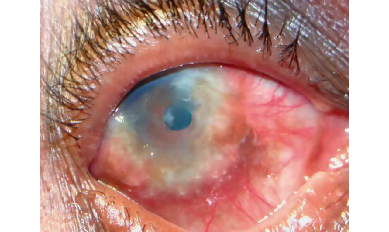
- Oedema: Diffuse sheet-like infiltration of conjunctiva may lead to pale white oedema and obscuration of blood vessels. Bulbar conjunctiva typically displays diffuse chemosis and injection.
- Conjuctival scarring: Conjuctival scarring may occur, often in reticular or septal pattern.
- Symblepharon (adhesion between palpebral and bulbar conjunctiva) formation: Sub-epithelial fibrosis can, in severe cases, lead to symblepharon formation.
- Forniceal foreshortening: Forniceal foreshortening may also result from sub-epithelial fibrosis.
- Limbus: Limbus may become infiltrated and oedematous, and develop gelatinous limbal thickening, consisting of confluent macro-papillae, is sometimes seen.
- Trantas dots: Trantas dots, tiny white lesions consisting of necrotic epithelial cells and eosinophils, may be observed, similar to those in VKC.
Cornea:
Corneal disease may be complicated by late development of corneal hypaesthesia in patients with AKC, resulting in a paradoxic reduction in surface symptoms, including itching.
- Punctate epithelial keratopathy: Punctate epithelial keratopathy is the most common corneal finding.
- Filamentary keratitis: Filamentary keratitis may occur, sometimes with very thick mucoid filamentary strands and possibly related to tear film instability, due to goblet cell abnormalities and deficient mucin secretion, also commonly featured in AKC.
- Persistent epithelial defects: Persistent epithelial defects frequently occur in the setting of a dry and inflamed ocular surface and may eventually lead to macro-erosions. Macro-erosions in atopic patient may progress to frank bacterial ulcers.
- Atopic shield ulcers with ‘vernal’ plaque formation: Atopic shield ulcers with ‘vernal’ plaque formation may also develop. Adherent mucus plaques contain epithelial debris, eosinophils, and inflammatory cells, and probably result from a combination of mechanical irritation from giant papillae, as well as toxic epithelial changes secondary to inflammation. Persistence of these plaques may eventually cause stromal thinning and perforation. The chronic inflammation and mechanical insult from palpebral scarring may result in partial or total limbal stem cell deficiency. Chronic inflammation and superimposed infection may lead to corneal scarring, neovascularisation, and lipid deposition. Vision may decline due to obscuration of the visual axis, irregular astigmatism, and/or ocular surface compromise.
- Pannus formation: Severe pannus often develops, typically affecting the superior one-third of the cornea.
- Pseudogerontoxon: Pseudogerontoxon may be seen in the peripheral cornea, which resembles a short, circumferential segment of arcus-senilis (white or grey opaque ring in corneal margin). This localised area of lipid deposition, related to abnormal vascular permeability at the limbus, may be the only evidence of previous atopic disease in a quiet eye.
Other complications/features:
- Eyelid inflammation: Eyelid inflammation is common in AKC, often related to staphylococcal blepharitis. Patients with atopic dermatitis are found to have high rates of bacterial skin colonisation, specifically with staphylococcal species.
- Corneal super-infections: AKC patients are at higher risk of corneal super-infections because of an unstable ocular surface, the local bacterial colonisation of the eyelids, and a dysfunctional innate immune system. Herpes simplex virus (HSV) keratitis, frequently bilateral, is another well known complication of AKC, and is presumably related to abnormalities in the atopic host’s immune defences. Herpetic epithelial lesions may be recurrent, especially when topical or systemic immune-suppressants are required to control the atopic state. Management is especially difficult because epithelial AKC lesions may be difficult to distinguish from HSV keratitis.
- Development of cataract: Rapidly progressive cataracts frequently develop in AKC patients, classically described as anterior sub-capsular opacities, usually stellate or shield like in appearance. The pathogenesis for atopic cataract is unclear, and it is suggested that high levels of IgE may be correlated with development of cataract in these patients. The chronic use of topical steroids also predisposes to posterior sub-capsular cataracts in AKC patients. Other forms of cataract may form independent of corticosteroid use, especially in patients with severe systemic atopic disease.
- Keratoconus: A higher incidence of keratoconus is reported in AKC, likely related in part to chronic eye rubbing.
- Pellucid marginal degeneration: A higher incidence of pellucid marginal degeneration is reported in AKC, likely related in part to chronic eye rubbing.
- Retinal detachment: Slightly higher rate of retinal detachment has also been noted in AKC patients. This may also be related to chronic eye rubbing, inducing degenerative vitreous changes.
Histological/ Immuno-histochemical studies:
Histological/ Immuno-histochemical studies of conjunctival specimens reveal increased number of mast cells, eosinophils, T lymphocytes and neutrophils in AKC patients.
Mast cells and eosinophils have not only been detected in conjunctival epithelium, but are also present in increased numbers in the substantia propria. A surplus of Langerhans cells, macrophages and B cells in the substantia propria is also seen.
T-lymphocyte infiltration is apparent in both conjunctival epithelium and substantia propria in AKC. T-helper (Th) cells predominate in all allergic eye diseases. Of the major T-cell subtypes, the Th1 subtype is involved in cellular immunity, whereas Th2 subtype is involved in humoral immune responses, including those mediated by IgE. Th2 cells are involved in eosinophil recruitment and stimulate IgE production from B cells. Th2 cytokines are involved in the recruitment and activation of inflammatory cells in AKC.
Activated eosinophils recruit additional inflammatory cells to the ocular surface. They release chemo-attractants which attract neutrophils to the conjunctiva. Eosinophils are also major players in the development of sight-threatening corneal complications in AKC.
Conjunctival Epithelial cells and Fibroblasts express pro-inflammatory mediators in VKC. The expression of surface adhesion molecules and the release of cytokines by epithelial cells enhance recruitment of eosinophils. Conjunctival fibroblasts, when activated by Th cytokines, promote local accumulation of eosinophils and enhance eosinophil degranulation.
Secondary structural changes:
Extensive inflammation in AKC leads to chronic ocular surface alterations:
- Goblet cell loss/hyperplasia: Both goblet cell loss (based on impression cytology), as well as goblet cell hyperplasia (based on conjunctival biopsy) have been reported. Goblet cell production of mucin is altered.
- Tear film instability: Tear film instability, with increased tear break-up time (BUT) but normal tear production, has also been described.
- Corneal sensation: Corneal sensations are reduced in AKC..
-Squamous metaplasia: Squamous metaplasia has also been described in patients with AKC.
Confocal microscopy studies:
AKC is associated with alterations in basal epithelium and sub-basal corneal nerves which relate to changes in tear function and corneal sensitivity. There is lower density of basal epithelial cells. A decrease in number and density of sub-basal long nerve fibers causes decreased corneal sensation. Corneal nerves are thickened and have bifurcation abnormalities. Inflammatory cells and activated keratocytes are found in the superficial stroma.
AKC should be differentiated from:
- Chronic allergic conjunctivitis (e.g. VKC).
- Blepharitis.
- Viral conjunctivitis.
- Trachoma.
- Cicatricial pemphigoid.
Similarities and differentiation from VKC:
Similarities:
- Potentially blinding chronic disease: AKC and VKC are chronic, potentially blinding conditions.
- Atopy: Both conditions present in individuals predisposed by an atopic background.
- Hypersensitivity responses: Both diseases involve both type I and type IV hypersensitivity responses. Eosinophils and T lymphocytes are found to infiltrate the conjunctiva in both conditions.
- Clinical features: Clinically, they are similar, with conjunctival inflammation, epithelial defects, shield ulcers, and corneal scarring as major features of both.
Differences:
- Age groups: AKC presents at an older age than VKC. VKC usually ‘burns out’ by late puberty, whereas AKC remains chronic for years, often persisting into old age, when it may resolve spontaneously.
- Visual prognosis: Visual prognosis is worse for AKC.
- Peri-orbital skin and eyelid: There is increased peri-orbital and eyelid skin involvement in AKC as compared to VKC which generally spare skin.
- Papillae: AKC usually presents with micro-papillae on the palpebral conjunctiva. Lower tarsus is preferentially involved in AKC, in contrast to VKC, in which papillae are much more prominent on the upper tarsus. Giant papillae are sometimes seen in AKC, but this is a definitive hallmark of VKC.
- Eyelid margins: The eyelid margins are affected in AKC unlike VKC. Cicatricial changes, forniceal foreshortening, and symblepharon may be seen in AKC which are not usual in VKC.
- Trantas dots: Trantas dots are more frequently associated with VKC than AKC.
Atopic Keratoconjunctivitis Complications
- Ptosis.
- Tylosis (thickening of eyelid margin).
- Staphylococcal blepharitis.
- Conjunctival fornix foreshortening.
- Keratopathy.
- Keratoconus.
- Recurrent herpes simplex keratitis.
- Corneal scarring.
- Corneal thinning.
- Corneal perforation.
- Pellucid marginal degeneration.
- Anterior sub-capsular cataract.
- Posterior sub-capsular cataract.
- Degenerative vitreous changes.
- Retinal detachment.
Atopic Keratoconjunctivitis Prevention
For optimal long term prevention of AKC, reduce or eliminate the exposure to environmental allergen.
Mast cell stabilisers and antihistamines are the mainstay of prophylactic therapy. Reduction of environmental allergens along with oral and topical antihistamines helps in management of exacerbations.