What is Conjunctival Concretions ?
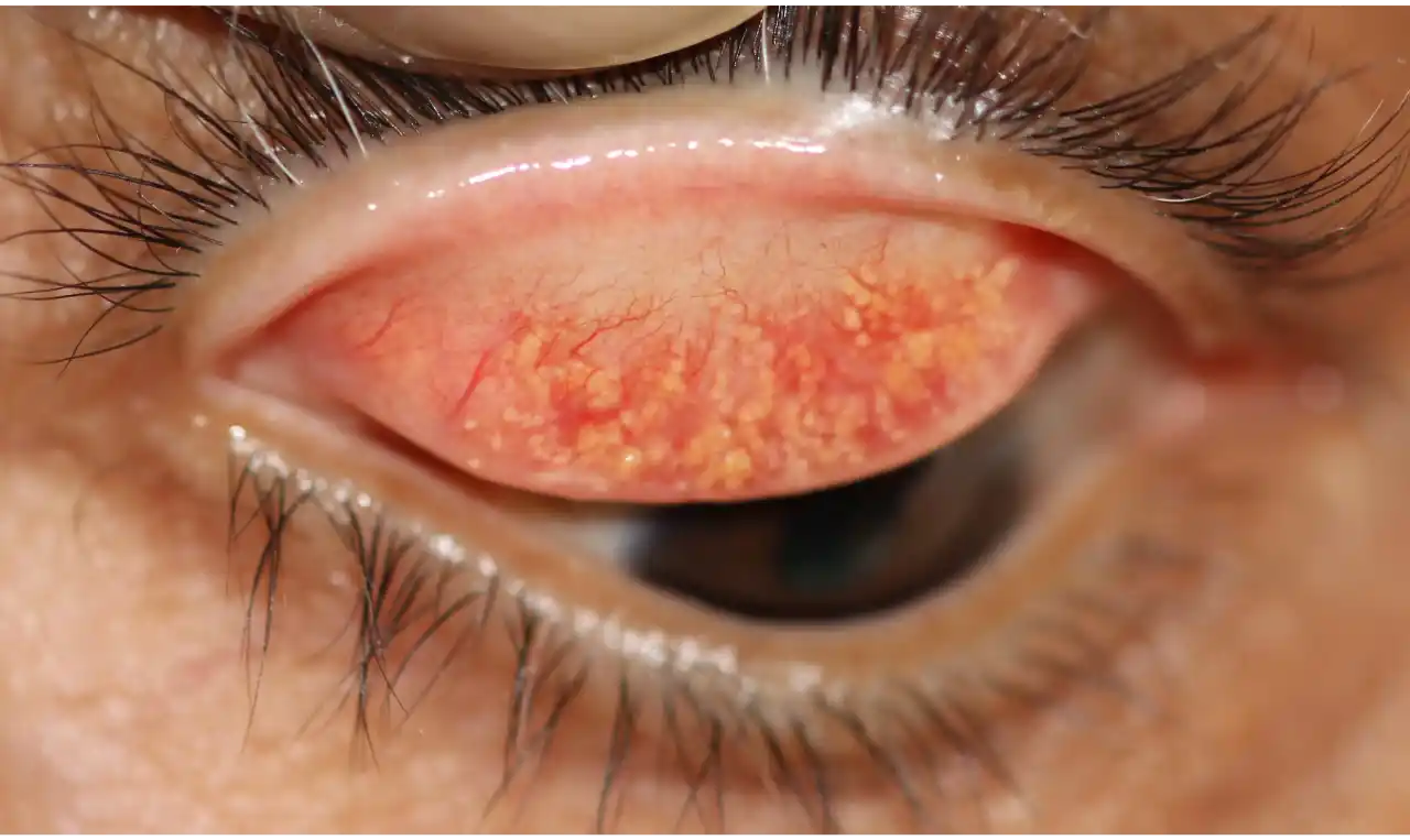
Conjunctival concretions (Conjunctival lithiasis) are small vascular, granular, yellowish-white deposits being produced due to conjunctival degeneration and are found in the sub-epithelium of palpebral conjunctiva and fornix (junction between palpebral and bulbar conjunctiva) in the elderly group or in patients with chronic inflammatory conditions. Deposits were found in patients as young as 20 years old, with the range of occurrence from 30-80 years old. Concretions appear as multiple tiny inclusion cysts containing yellowish-white deposits of inspissated mucous and degenerated epithelial debris including keratin. They are frequently discrete, but confluent concretions are not uncommon. In one study, no significant difference was found between the lacalisation on the upper and lower eyelids, right or left eye, and most of the concretions were superficial and hard, and mainly single. Associated dysfunction of meibomian glands was also noticed in some patients.
A recent article has emphasised how patients with conjunctival concretions are potentially affected with dry eye disease.
They are usually idiopathic, but they have also been associated with:
- Chronic atopic keratoconjunctivitis.
- Following post-trachomatous degeneration.
- Sulphadiazine eye drop administration.
Degenerations of the conjunctiva are common conditions that, in most cases, have relatively little effect on ocular function and vision. These conditions increase in prevalence with increasing age as a result of past inflammation, of long-term toxic effects of environmental exposure causing deposition, or of ageing itself. Conjunctival degenerations may be associated with chronic irritation, dryness, or previous history of trauma.
Identification of corneal and conjunctival degenerations has improved with the use of confocal microscopy, immuno-histochemical staining, and genetic testing.
Conjunctival Concretions Symptoms
Concretions are almost always asymptomatic because they usually remain hidden into the palpebral conjunctiva, not noticed by patients, until they become large and protrude through the palpebral tissues.
Concretions may irritate eyes, but only if they erode the overlying conjunctival epithelium and come in contact with the cornea.
Protruding concretions may cause symptoms such as:
- Foreign body sensation.
- Lacrimation (watering).
- Discomfort.
- Irritation.
- Redness.
- Corneal abrasion (less common).
Conjunctival Concretions Causes
Concretions are formed due to the accumulation of epithelial cells and inspissated mucous in depressions called Henle glands.
The causes and risk factors for conjunctival concretions are variable but are associated most commonly with ageing and chronic inflammation of conjunctiva.
Causes and risk factors are:
- Ageing process.
- Chronic conjunctival inflammation (e.g. trachoma).
- Tear film deficiency (present as decreased values of the Schirmer’s test and tear film break-up time).
- Severe atopic keratoconjunctivitis.
- Meibomian gland dysfunction (e. g. chronic meibomitis).
- Re-crystallisation of certain eye drops (e.g. sulphadiazine)
Conjunctival Concretions Diagnosis
Diagnosis of conjunctival concretions is often incidental. Patients are usually asymptomatic and conjunctival concretions are noted on ocular examination. Some patients with protruding concretions may complain of irritation and foreign body sensation in eye. Patients may or may not give history of chronic conjunctivitis.
Clinical examination:
- Conjunctival concretions are small 1- 2 mm, yellow-white lesions typically present on palpebral conjunctiva and fornices (plural of fornix).
- Concretions may be single, multiple, or confluent rarely.
Histology and Electron Microscopy:
The primary components of conjunctival concretions are degenerating epithelial cells and inspissated secretions from conjunctival glands. Following inflammation, the debris get trapped in sub-conjunctival depressions (Henle glands), and they often undergo calcification. The histology shows mucinous secretion from conjunctival glands. Occasionally, epithelial cysts are located on top of the concretions.
Conjunctival concretions are finely granular and membranous debris. These concretions were thought to be calcifications, thus the misnomer ‘conjunctival lithiasis’.
Staining properties:
- Strong staining: In contrast to calcium staining, concretions stain strongly for phospholipid and elastin.
- Weak staining: Concretions stain weakly for neutral polysaccharides and lipids.
- No staining: Concretions stain negative for amyloid, collagen, glycogen, iron, muco-polysaccharides, calcium, RNA, and DNA.
Electron Microscopy:
Although conjunctival concretions may have substantial calcification, they are not typically calcareous hard and do not show crystalline pattern under electron microscope, therefore, the term lithiasis should not be applied.
Conjunctival concretions should be differentiated from:
- Epidermal inclusion cysts.
- Lymphoid follicles.