What is Amblyopia ?
Amblyopia is often defined as a difference in visual acuity of two lines or more on Snellen or equivalent chart in a child with, otherwise, healthy eyes. Amblyopia may be present any time visual acuity is reduced, and the reduction of acuity cannot be explained by findings on clinical examination, even if the difference is even one line only.
The term amblyopia is derived from Greek word (amblys means dull and ops means eye), which literally means dullness of vision. Von Noorden defined amblyopia as a ‘decrease of visual acuity in one eye when caused by abnormal binocular interaction or occurring in one or both eyes as a result of patterned vision deprivation during immaturity, for which no cause can be detected during the physical examination of the eye(s) at which in appropriate cases is reversible by therapeutic measures’.
Amblyopia is a functional reduction in the visual acuity of an eye caused by disuse during critical period of visual development. The mechanism of vision loss is not known, but it is thought to originate in the visual cortex. Amblyopia results in reduced visual acuity, binocularity, depth perception, and contrast sensitivity. Fusion and stereopsis, the central formation of three dimensional images, are dependent upon receiving clear images from each eye simultaneously
Amblyopia Symptoms
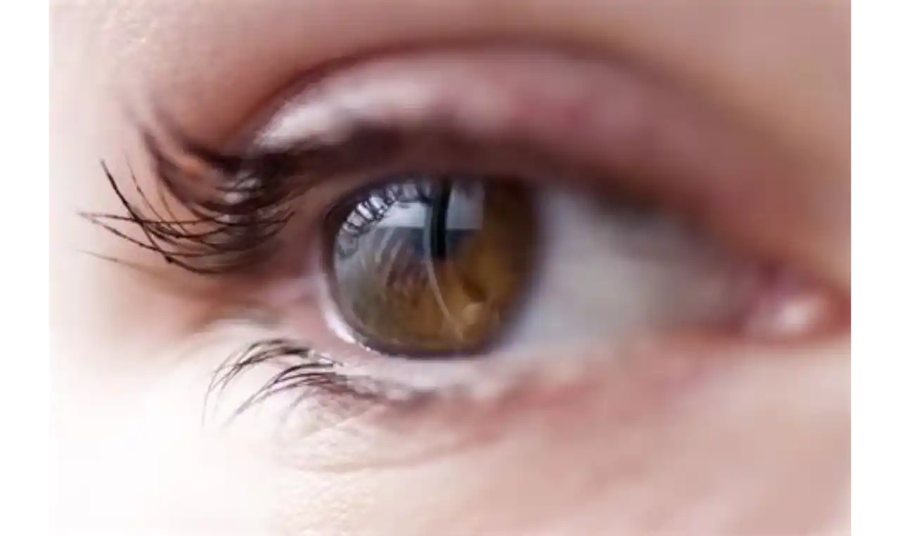
The main symptom of amblyopia is decreased foveal visual acuity. Commonly used diagnostic criteria, is the loss of visual acuity of two or more lines on the Snellen vision chart.
- Amblyopic eye presents an abnormal contour interaction which shows decrease in acuity for objects placed in a row compared with acuity for the same objects viewed separately (crowding phenomena).
- Eccentric fixation.
- Decreased contrast sensitivity.
- Improved vision in dim background illumination.
- Decreased brightness perception.
- Binocular suppression of amblyopic eye.
Amblyopia Causes
A distinction must be made between potentially reversible functional amblyopia and irreversible organic amblyopia. Organic amblyopia is a term used to describe visual impairment due to obvious or non obvious ocular pathology, commonly affecting retina or optic nerve. Examples of organic amblyopia are optic nerve hypoplasia, optic atrophy and foveal hypoplasia. Functional amblyopia may occur along with organic amblyopia. Functional amblyopia normally occurs in an eye that is anatomically normal.
Functional amblyopia is caused by either form of vision deprivation or abnormal binocular interaction. Form- vision deprivation occurs due to conditions that obstruct the visual axis such as cataract, corneal opacity, vitreous haemorrhage, or severe ptosis, but it may also be produced by severe anisometropia. Abnormal binocular interaction refers to the condition in which the image projected onto the fovea of each eye is dissimilar enough to preclude fusion, thus prompting suppression and ultimately amblyopia of the suppressed eye. While strabismus may be the most obvious cause of abnormal binocular interaction, unilateral opacity of the media and anisometropia may participate in this mechanism as well.
Aetiological classification: This is the most commonly used classification and has high prognostic value. It is classified as
- Strabismic amblyopia: Frequently amblyopia is associated with strabismus. Often it may not be known with certainty whether amblyopia is the consequence or the cause of strabismus. Constant tropias (squint) are most commonly associated with amblyopia unlike intermittent tropias (intermittent exotropia). This type of amblyopia is usually unilateral and the severity is not related with the degree of strabismus.
- Refractive amblyopia: Ametropia is one of the most important causes of uni- and bilateral amblyopia.
- Isoametropic amblyopia: This is related to the age. It is said that high refractive errors either bilateral or unilateral would not affect the development of binocularity and visual acuity before the first year of life. Otherwise, refractive errors not corrected after one year of age are related with a high degree of uni- and bilateral decrease of vision.
In hypermetropia, insufficient accommodative effort which inadequately focuses the images on retina may produce amblyopia. In high myopia, objects close to the patient tend to be in focus and amblyopia is uncommon.
- Anisometropic amblyopia: It is likely to occur because the amount of accommodation necessary to focus the image in the less hypermetropic eye is utilised. Therefore, more hypermetropic eye receives blurred image. When the difference in hypermetropia exceeds two dioptres, amblyopia may be present. Some cases even with less amount of difference may develop amblyopia.
In anisomyopia, more myopic eye may be used for near vision and the less myopic for distance vision. It causes sharp image to be present in both the eyes. Amblyopia is unusual when myopia does not exceed five dioptres.
- Meridional amblyopia: Meridional amblyopia is due to astigmatism producing defocused images with specific orientation. Astigmatism appears to be an important factor for amblyopia, during first year of life.
- Stimulus deprivation amblyopia: This produces most severe amblyopia. Nystagmus usually presents with amblyopia during first three months of life. Ptosis, congenital cataract, corneal opacity and eye occlusion also produces stimulus deprivation amblyopia. Unilateral cases are more amblyogenic. In general, early the treatment, better is the result. Congenital cataract is the leading cause of deprivation amblyopia and need quick diagnosis.
- Idiopathic amblyopia: In some patients no amblyogenic factor is detected. It is postulated that this may be due to some amblyogenic factor, such as transient anisometropia in infancy, which might have disappeared with advancing age.
- Occlusion amblyopia: Occlusion amblyopia is an iatrogenic condition caused by therapeutic patching of the eye with normal vision. It usually occurs in the sound eye as a result of amblyopia treatment of other eye, but it is occasionally seen in young children after occlusion therapy to treat ocular pathology, such as corneal abrasion.
Amblyopia Diagnosis
Diagnosis of amblyopia is based mainly on measurement of visual acuity.
A diagnosis of amblyopia requires complete eye examination to rule out any organic cause as well as correct refractive error under cycloplegia. Clinically an organic cause is suspected in addition to functional amblyopia, if treatment improves vision up to certain level only. Amblyopia may be mimicked by any defect in the afferent visual system. Optic nerve glioma is the most important suspected pathology.
Visual acuity measurement
Subjective and objective methods to test visual acuity have been proposed.
- Optical grating: The different stimuli consist of repetitive gratings of predictable and precise luminance variations. Sinusoidal and square- wave gratings are some other forms to test visual acuity.
- Optokinetic nystagmus: The primary use of optokinetic nystagmus is a rapid screen for the gross integrity of the visual system. Acuity may be measured as the finest grating that elicits a visible optokinetic nystagmus when different sizes of stripes are used.
- Preferential looking test: It is based on the principle that infants tend to fixate a pattern stimulus rather than a homogeneous field. With this, inter-ocular acuity may be detected in children less than one year of age. Acuity is estimated by selecting the spatial frequency of the stripe that was fixed longer than the homogeneous field by 75% of the infants at a given age.
- Visually evoked potential: Visually evoked potential is a summed cortical response to changes in some characteristics of a visual stimulus. The visual stimulus may be either a simple flash or a more complex pattern. All these techniques are complementary to evaluate vision in children who are unable to complete recognition acuity tests.
- Visuscope: Visuscope is a type of direct ophthalmoscope that projects a focused image onto the retina so that the examiner can see the image on the retina. First the image is projected onto the parafoveal retina, and then the patient is asked to look at the image. Patient with central fixation re-fixates to place the image on fovea again. However, with eccentric fixation, the patient views with the parafoveal retinal area and show a wandering unsteady fixation.